In The Anatomy of Bees, we took a look at the body parts of the honeybee and the impressive efficiency of this marvel of nature. In this lesson we drill down a little further, to explore some other details of this remarkable creature.
The Head
Inside and out, a bee’s head is focused on sensing its surroundings and responding to what it senses.
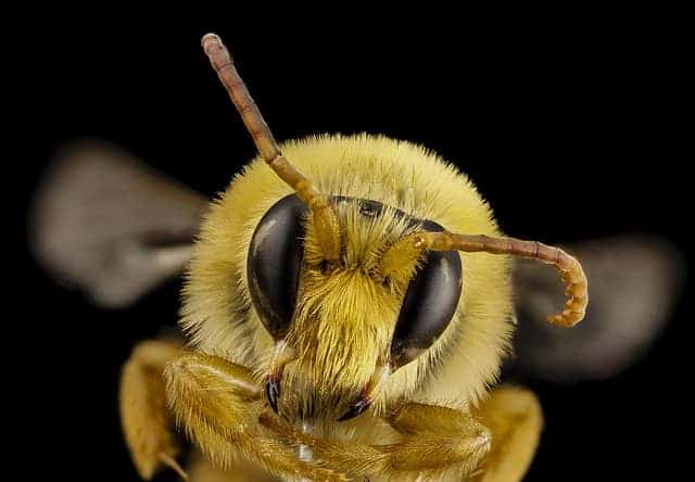
Four appendages are located on the bee’s head – the antennae, the mandibles, the maxillae and the labium. The compound eyes and ocelli are also located on the head.
Antennae
Each antenna is attached to the head with a ball and socket. This antennae base is called a scape. The antennae culminate with flagella. The flagella and scape are connected by a flexible elbow call the pedicel.
Each antenna is covered with hairs and thousands of other sensory structures. All these structures are connected with nerves that send messages to the brain and other parts of the nerve system. These sensory structures can be divided into:
- Odor receptors (or smell sensors)
- Gustatory receptors (or taste sensors)
- Mechanoreceptors (or touch sensors)
- Proprioceptors (or position sensors)
At the base of the antenna is the Johnston’s Organ. The sensory receptors on the flagella sense the movement of air and this movement is relayed to the Johnston’s Organs, allowing a bee to hear. These messages also allow a honey bee to tell how fast and far it is flying.
The Johnston’s organ is essential in receiving communication from scout bees. They do this during their dance communicating the location of resources, as well as signaling the location of a new nest sight when swarming. It is thought that the movement of the wings of scout bees creates an electrical field that is received by the Johnston’s Organ.
Mandibles
The mandibles are suspended from the side of the mouth and help to protect the mouth parts. The primary purpose of mandibles is for working with wax, but they do have other purposes.
Mandibles can be used to bite a flower petal, allowing a bee to access nectar. They can even be used to chew wood, but since the muscles powering the mandibles are very weak, this is not a frequent task honey bees undertake.
Honey bees do use the mandibles to bite. A person would barely feel this bite, but to a wasp or to varroa mites, a bite from the mandibles can be fatal.
At the base of the mandibles, a tube runs to the mandibular gland. Mandibular glands are present in all honey bees, but are largest and most functional in the queen. In the queen, these glands produce the queen pheromone or queen substance. This pheromone is the major factor in maintaining the cohesiveness of the hive.
In young worker bees, the mandibular glands produce a substance that is mixed with secretions from the hypopharyngeal glands to make royal jelly.
The mandibular glands also secrete 2-heptanone. This same chemical is produced in the sting apparatus. The purpose of 2-heptanone is not fully known. We do know that 2-heptanone produces a banana smell that is associated with a bee signaling other bees to attack. It is also believed that 2-heptanone has anesthetic properties allowing honey bees to bite pests, stunning them long enough for them to be removed from the hive.
Maxillae and Labium
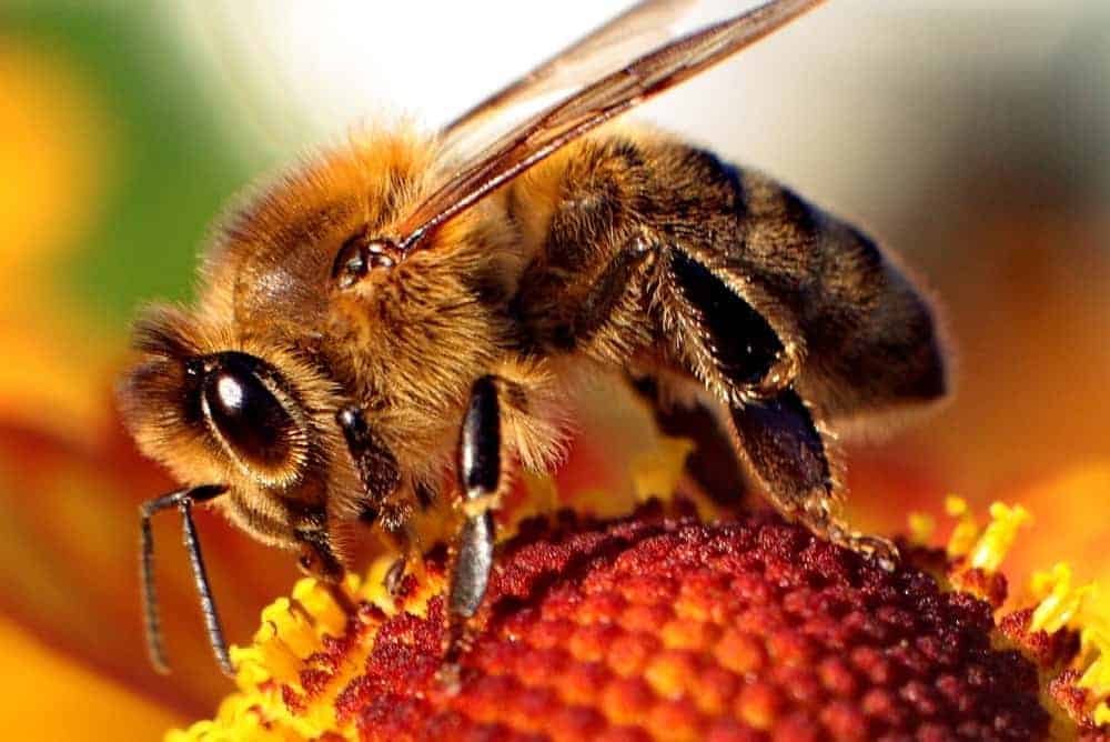
In most insects, the proboscis is a permanently functioning organ, but in a honey bee, the proboscis is temporarily brought together by joining the maxilla and labium. This allows a honey bee to suck liquid. The maxillae and labium are then separated allowing the bee to eat solid food.
The sucking ability of the proboscis is powered by a sucking pump in the head. This pump moves liquid from the proboscis to the esophagus. It also enables a bee to move liquids in both directions, allowing a worker to regurgitate liquid once it is back in the hive.
Eyes
The larger part of a honey bee’s head is composed of eyes. There are two types of eyes.
Three simple eyes, called ocelli, form a triangle at the top of a bee’s head. The light they sense is essential in determining the position of the sun and guiding navigation. Each ocellus has a single lens positioned over 800 light receptor cells. No image is formed from the light received by the ocelli. It is believed that one purpose of these eyes is to inform the honey bee when it is time to begin foraging and when it is time to quit for the day.
The two compound eyes in worker bees are composed of hexagonal facets. Groups of facets cover retinal units. A retinal unit is called an ommatidia. Each ommatidia has its own lens and a bundle of eight or nine photoreceptor cells.
Each ommatidia receives a different picture of the field of view. A worker bee has approximately 6,900 ommatidia. Drones have approximately twice that number of ommatidia.
A bee’s vision is somewhat like a pixilated TV picture. It is estimated that the visual acuity of honey bees is about one percent that of humans. Like humans, honey bee vision is trichromatic. Where humans base their vision on red, blue and green, honey bee vision is based on blue, green and UV. This means that honey bees cannot see red. A field of red poppies will appear as black blobs to bees.
The Brain
The largest organ inside the head is the bee’s brain. The hypopharyngeal gland and the labial glands are also found inside the head.
Honey bee brains have an optical lobe which receives input from the compound eyes and an antennal lobe that receives input from the antennae. Between these two lobes is a section called the “mushroom body”. A cross section resembles two mushrooms. This section is involved with olfactory learning and memory.
Instructions from the brain for bodily functions is communicated via seven ventral ganglia (nerve cell bundles) running through the thorax and abdomen. Nerve cells run from each ganglion to the organs regulating the functions of each organ.
If you have some time, check out this video and learn something new about the honey bee brain!
The Hypopharyngeal Gland
This gland consists of a pair of tubes coiled against the sides of the head, between the front cuticle and the brain. It secretes a protein substance called royal jelly or bee milk. Worker and drone brood receive this royal jelly for the first two or three days of their lives. Queens receive royal jelly their entire larval lives.
A honey bee is only efficient in producing royal jelly for a few days. In older forager bees, this gland begins to produce an enzyme that breaks sucrose into fructose and glucose.
Salivary Glands or Labial Glands
The salivary glands are also called labial glands. There are two sets of salivary glands. One set is at the rear of the head and the other in the thorax, just behind the head.
The salivary glands produce saliva used to dilute food and dissolve sugar. These glands also produce compounds used to clean the body and chemicals that give the hive its identity. Saliva produced in the glands located in the head is mixed with wax scales to shape wax.
Thorax
The thorax is composed of three segments: the prothorax, the mesothorax, and the metathorax. Each segment has one pair of legs. The back two segments each have one pair of wings.
The main components inside the thorax are muscles and nerves. The nerve center of the thorax controls the muscles. The muscles in the thorax control the legs, wings, head movement and abdomen movement.
The Legs
The most obvious function of a bee’s legs is locomotion. The legs function in a triangle with the first and third leg on either side working with the middle leg on the opposite side.
There are, though, other less obvious functions of the legs. A bee’s legs are vital for communication. Subgenual organs in the lower part of the legs allow a bee to pick up vibrations created by scout bees as they perform their waggle dance to communicate the location of food.
Legs also have sensory organs that taste, smell and feel. The first pair of legs have special tools for cleaning antennae. The middle legs have stiff spines that are thought to be used to spear wax scales and to pass them to the bee’s mouth. The third pair of legs have special stiff hairs, arranged like a comb, for cleaning pollen from body hairs.
The Wings
In addition to flying, wings are vital for regulating temperature and moisture in the hive. A bee can disengage its wings from the wing muscles to allow it to use its body as a heater. Flight muscles can contract several times with one single nerve impulse. This allows for the wings to beat up to 230 times per second.
Abdomen
The abdomen consists of ten segments, with only seven visible. The abdomen has no outside structures, but houses the honey crop, the wax glands, the scent gland, digestive system and the sting.
Honey Crop
Once a bee collects nectar or water, that nectar moves through the head region and the thorax into the abdomen where it enters the crop. When full, the crop occupies almost half the abdomen.
The preventriculus closes the crop to prevent its contents from entering the digestive system and opens to allow content to move to the digestive system. The content of the crop is regurgitated once the worker is back in the hive.
Wax Glands
There are four wax glands. Worker bees develop the ability to produce wax once they are between 6 and 12 days old. Honey bees are only efficient at wax production for about 6 days. After about 6 days of wax production, the wax glands deteriorate.
Nasonov (Scent) Gland
The Nasonov, or sent gland, produces Nasonov scent pheromone. This pheromone is used by honey bees to assist in orienting. It is mainly used to identify water sites and to identify the location of a new hive. Maximum ability to release the Nasonov pheromone is developed in forager bees.
Digestive System
Food that passes through the crop enters the midgut or ventriculus. This is the functional stomach of a bee. Malpighian tubules, small tubes near the end of the ventriculus, remove waste and pass it to the rectum. The rectum is expandable and allows a bee to retain solid waste for months during the winter.
Undigested material is passed through the anus, often while in flight. Most liquid and nutrients are removed before passing through the anus.
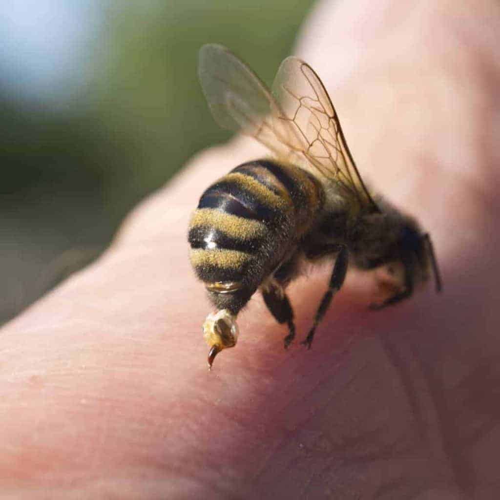
The Sting
There are at least four glands associated with the sting of a honey bee. These glands produce an alarm pheromone, venom for the sting and histamine that is injected with the sting.
The sting itself is designed in such a way that it easily pulls out of other insects, but barbs on the sting prevent it from easily pulling out of the skin of people. When a bee attempts to flee after stinging, the sting, the venom gland and the muscles controlling the gland break away from the bee. The muscles continue to pump venom into the victim. The alarm pheromone is also released. This alarm pheromone signals other bees to sting.
Summary
The honeybee is an incredible living bundle, packing an impressive series of capabilities into a tiny frame. When this little powerhouse is aggregated across the tens of thousands of bees in each colony, we start to understand how the productivity of our hives rivals anything seen in nature.
Author : William Gullick, PerfectBee Ambassador,

Sources
Bee Health Extension. (2019). Adult Bee Anatomy (Basic Bee Biology for Beekeepers).
Ask a Biologist. (n.d). Bee Bonanza: The Story of Honey Bees.
Caron, Dewey M. & Connor, John Lawrence. (2013). Honey Bee Biology and Beekeeping. Kalamazoo: Wicwas Press.
Graham, Joe M. (2015).The Hive and the Honey Bee. Hamilton, Illinois: Dadant and Sons.
Snodgrass, R. E. (1910). The Anatomy of the Honey Bee. Entomology Bureau. Technical Series. No. 18. 162pp. Washington, D.C. Government Printing Office.
Tautz, Jürgen. (2009). The Buzz about Bees: Biology of a Superorganism. Heidelberg: Springer.